Brian Hudelson, University of Wisconsin Plant Disease Diagnostics Clinic
When I was asked to write this article on diagnosing zonate leaf spot (caused by various species of Cristulariella/Grovesinia), I was a bit reticent. I am in no way an expert on this disease or the pathogens involved. In fact, I have only seen zonate leaf spot three times in my 25 years at the UW-PDDC. Interestingly, the first two samples arrived on the same day in 2018, from two somewhat geographically distant Wisconsin counties, and on different hosts (maple and grape). I subsequently saw the disease again in 2019 on industrial hemp.
In my initial encounter with zonate leaf spot, a UW-Extension county educator sent me photos of maple leaves with symptoms (roughly round lesions with a pattern of concentric rings) that were typical of what I had seen online for zonate leaf spot. I REALLY wanted to see a physical sample (and offered to do the diagnosis for free), as zonate leaf spot had been on my plant disease “bucket list” ever since I first saw drawings of reproductive structures (conidiomata) of Cristulariella in Barnett’s Illustrated Genera of Imperfect Fungi. The Extension educator was quite accommodating.
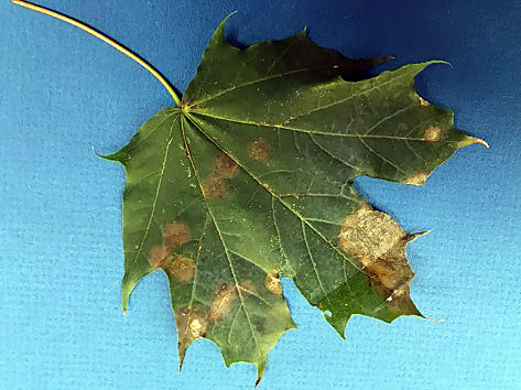
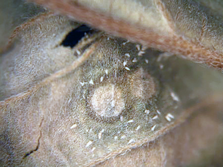
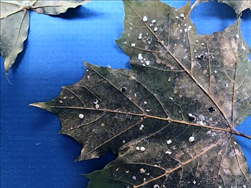
Once the physical sample arrived, I again noted typical zonate leaf spot lesions (Figure 1). I scanned the lesions under my dissecting microscope and relatively quickly found conidiomata of the pathogen. These reproductive structures are fairly large (as fungal reproductive structures go), multicellular, tannish, and pyramidal in shape (Figure 2). Quite frankly, they look like tiny, tan Christmas trees popping up from the leaf surface. If you are in the field, you should be able to readily see these structures with a 20X hand lens. I ended up incubating the maple leaves in a moist chamber for several days, hoping to stimulate production of additional conidiomata. Interestingly, the pathogen eventually formed black sclerotia on the leaves that were readily visible to the naked eye (Figure 3).
When I encountered zonate leaf spot again in 2019 on hemp, I decided to go a step further in the identification, as I was not able to find references for zonate leaf spot occurring on this host. To confirm the identity of the fungus, I plucked one of the conidiomata from the leaf surface using a flame-sterilized needle and placed it on the center of a plate of ¼ strength potato dextrose agar amended with 100 ppm streptomycin sulfate. The fungus grew quickly, and Sue Lueloff, the molecular diagnostician in my clinic at the time, was able to sequence the ITS region of the isolate and identify it as Grovesinia moricola.
All in all, I have found the diagnosis of zonate leaf spot pretty straightforward. Hopefully, you will too. If you have questions about my work with zonate leaf spot and Cristulariella/Grovesinia, feel free to contact me at hudelson@wisc.edu or (608) 262-2863.
Search form
P&V Tips: In the Zone: Identifying Zonate Leaf Spot
![]()
NPDN supports plant health and biosecurity in U.S. agricultural and natural ecosystems by providing expert diagnostic capacity, communication, coordination, and quality pest and disease diagnostic information.
Copyright © 2026. All rights reserved.